


Home / Training / Manuals / Atlas of breast cancer early detection / Cases
Atlas of breast cancer early detection
Go back to the list of case studies
.png) Click on the pictures to magnify and display the legends
Click on the pictures to magnify and display the legends
| Case number: | 047 |
| Age: | 67 |
| Clinical presentation: | Postmenopausal woman with past history of ovarian carcinoma and increased risk of developing breast cancer presented with a right breast lump. Examination revealed a diffuse hard lump in the right breast. |
Mammography:
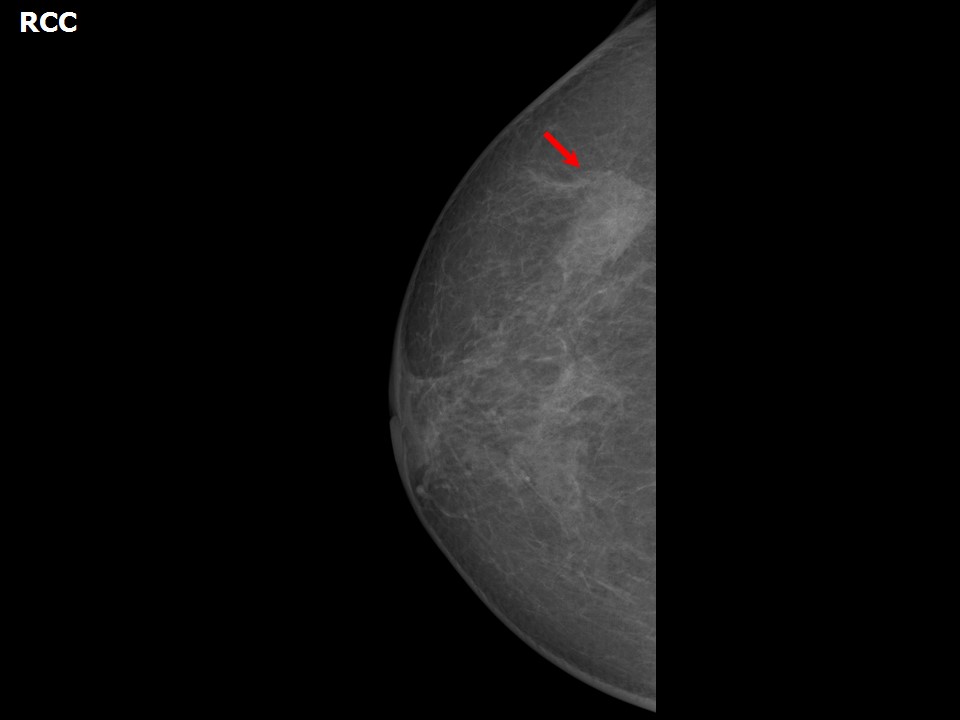 | 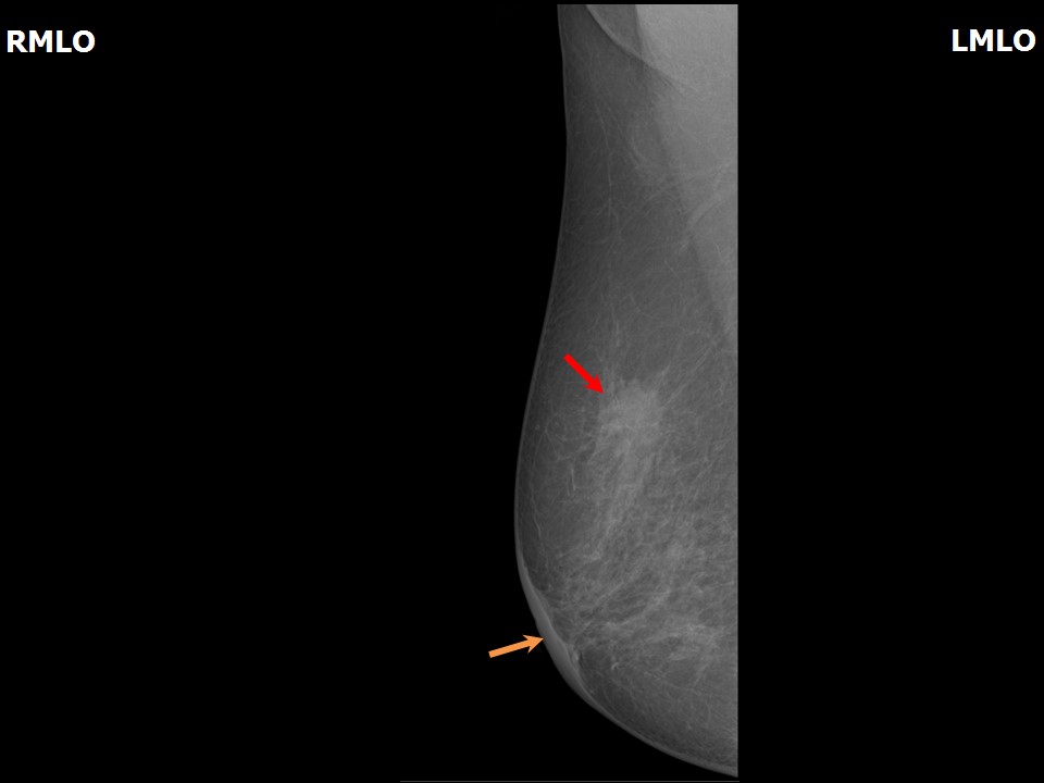 |
| Breast composition: | ACR category b (there are scattered areas of fibroglandular density) | Mammography features: |
| ‣ Location of the lesion: | Right breast, upper outer quadrant at 10 oclock, middle third |
| ‣ Mass: | |
| • Number: | 1 |
| • Size: | 2.5 × 1.7 cm |
| • Shape: | Irregular |
| • Margins: | Indistinct |
| • Density: | Equal |
| ‣ Calcifications: | |
| • Typically benign: | None |
| • Suspicious: | None |
| • Distribution: | None |
| ‣ Architectural distortion: | Present |
| ‣ Asymmetry: | None |
| ‣ Intramammary node: | None |
| ‣ Skin lesion: | None |
| ‣ Solitary dilated duct: | None |
| ‣ Associated features: | Trabecular thickening and skin thickening |
Ultrasound:
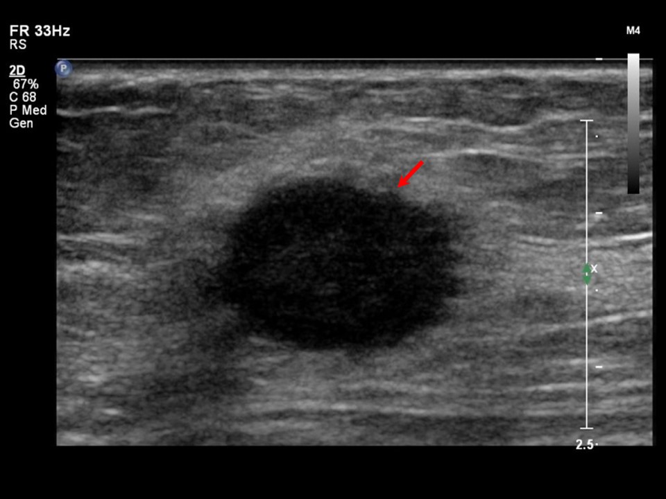 | 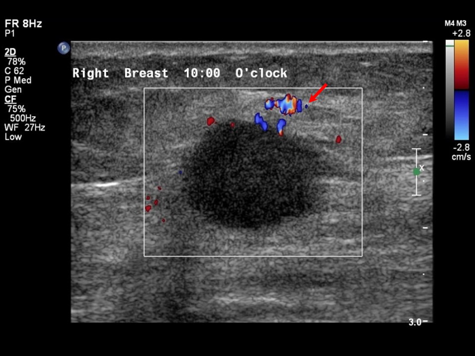 |
| Ultrasound features: Right breast, upper outer quadrant at 10 oclock | |
| ‣ Mass | |
| • Location: | Right breast, upper outer quadrant at 10 oclock |
| • Number: | 1 |
| • Size: | 2.0 × 1.5 × 1.2 cm |
| • Shape: | Irregular |
| • Orientation: | Parallel |
| • Margins: | Microlobulated |
| • Echo pattern: | Hypoechoic |
| • Posterior features: | No posterior features |
| ‣ Calcifications: | None |
| ‣ Associated features: | Vessels in rim around the mass |
| ‣ Special cases: | None |
BI-RADS:
BI-RADS Category: 5 (highly suggestive of malignancy)Further assessment:
Further assessment advised: Referral for cytologyCytology:
 |
| Cytology features: | |
| ‣ Type of sample: | FNAC |
| ‣ Site of biopsy: | |
| • Laterality: | Right |
| • Quadrant: | Upper outer |
| • Localization technique: | Palpation |
| • Nature of aspirate: | Whitish |
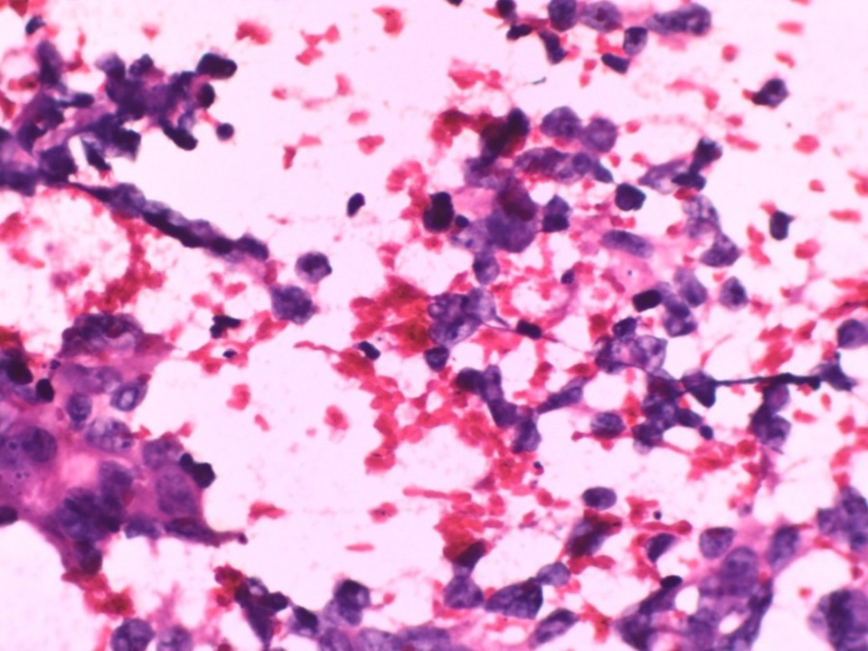
| ‣ Cytological description: | Smears are very cellular and show many dyscohesive sheets of malignant ductal cells. Single isolated malignant cells are also seen |
| ‣ Reporting category: | Malignant |
| ‣ Diagnosis: | Carcinoma high grade |
| ‣ Comments: | None |
Histopathology:
Breast-conserving surgery
 |
| Histopathology features: | |
| ‣ Specimen type: | Breast-conserving surgery |
| ‣ Laterality: | Right |
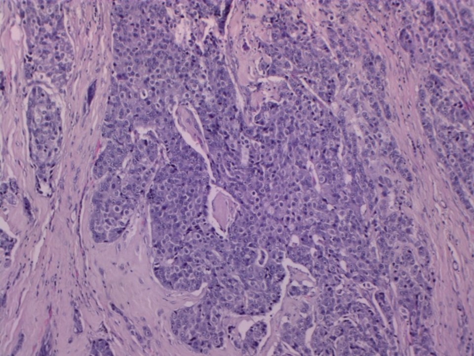
| ‣ Macroscopy: | Specimen (6.0 × 5.5 × 2.5 cm) with a strip of skin (3.0 × 0.8 cm) seen on anterior surface. Cut surface shows a tumour (2.5 × 2.5 × 1.5 cm) (T2) 2.0 cm from the lateral margin, 1.5 cm from the medial margin, 0.3 cm from the anterior margin (skin), 0.7 cm from the base (posterior margin), 1.0 cm from the superior margin, and 2.0 cm from the inferior margin. The cut surface of the tumour is greyish white with well-defined margins and it has a firm to hard consistency |
| ‣ Histological type: | Invasive breast carcinoma |
| ‣ Histological grade: | Grade 3 (3 + 3 + 2 = 8) |
| ‣ Mitosis: | 14 |
| ‣ Maximum invasive tumour size: | 2.5 cm in greatest dimension |
| ‣ Lymph node status: | 0/22 |
| ‣ Peritumoural lymphovascular invasion: | Absent |
| ‣ DCIS/EIC: | Absent |
| ‣ Margins: | Free of tumour |
| ‣ Pathological stage: | pT2N0 |
| ‣ Biomarkers: | |
| ‣ Comments: |
Case summary:
| Postmenopausal woman presented with right breast lump. Diagnosed as right breast carcinoma with areolar skin thickening, BI-RADS 5 on imaging, as right breast carcinoma on cytology, and as invasive breast carcinoma of no special type, pT2N0 on histopathology. |
Learning points:
|